
|
Saturday, July 22, 2006
I'm finally shutting down the old website, but I thought it might be nice to have some of these older posts to link to. Plus I'm moving to NYC and writing up a thing for my actual work, so not as much time for original writing for a month or so. This was my first ever blog post (with slight modification).
The title comes form Herbert Lubs, the first person to describe the chromosomal aberration for which Fragile X syndrome is named. The more extended quote goes "In one-third of the proband's metaphase figures, an unusual secondary constriction was seen at the ends of the long arm of a group C chromosome. This gave the appearance of large satellites, but the chromosome was never seen in association with the acrocentric chromosomes." 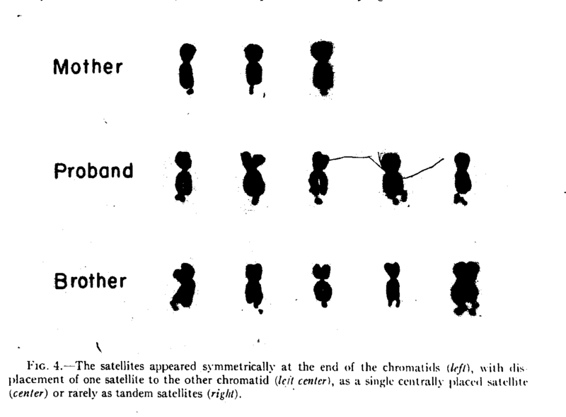 Part of the appeal of talking about Fragile X is that it covers so many levels of research. In this post, I'm going to give the basic features, a little history, and a little cytogenetics. I had planned on trying to explain why the Fragile X chromosome looks the way it does anyway, and it's fortuitous that there is an a blip on the chromosome radar coming up right as I prepare to do this. So without further ado, here's the litany of facts that you could've got if you'd only just googled Fragile X Syndrome:
Since I started off with the appearance of fragile X chromosomes I thought I'd try to explain why they look the way they do. This information was harder to find than I expected. It would appear that people are still working on this problem while I thought it would have been an almost immediate fallout from the discovery of the disease mechanism. Here's what I've got so far. "Microscopy" turned out to be a useful search term. First, there is a bit of an excuse to add a couple more pretty pictures. I found something of a methods paper describing a new way to mount chromosomes for transmission electron microscopy (TEM). These authors don't waste any time theorizing or introducing or giving background information. They want everyone to be aware of this new awesome technique and that's it. So it's kind of hard to understand, but the gist of it is that not only can you see the fragile site on these X chromosomes depicted as a reduction in electron density at the boundary between the main chromosome and the satellite, but you can also see other pockets of reduced electron density throughout the satellite. The picture below shows the tip of a normal chromosome on the left and a fragile one on the right. See how the fragile one is less dense? They can even see this pattern at least one chromosome that doesn't show a fragile sight when they use regular fluorescence light microscopy which may indicate a burgeoning fragile site. My understanding is that low electron density means that there are less proteins there. This is consistent with the story from this other paper. 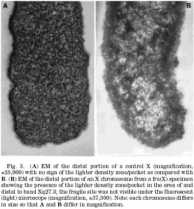 DNA during metaphase, which is when the pictures of chromosomes that you always see are taken, is coiled up and packed really tightly and part of the way this is achieved is to wrap the DNA strand around proteins complexes called nucleosomes. This is not the only case where DNA is wrapped around nucleosomes. It is pretty much happening all the time and DNA is getting wrapped and unwrapped all the time. Wang et al looked at the effect of having a bunch of CCGs repeated on this process. The way they did this was to mix small pieces of DNA (with or without a CCG repeats near one end) that were ostensibly not attached to any protein at the time with purified histones and let nature run its course. Then they took pictures with an electron microscope and counted places where nucleosomes had hooked up with the DNA. They found that while the distribution was fairly even across the non-CCG repeat DNA, there were hardly any nucleosomes at the location of the CCG repeats. So having a bunch of CCG's in a row makes it hard for nucleosomes to get involved. In fact, in later experiments, they found that nucleosome assembly was 62% less effective when there was a set of 76 repeated CCG's. They suggest that this leaves the DNA vulnerable to a process called methylation, by which one of those C's basically gets an extra carbon molecule sticking off of it that can alter the way the DNA interacts with proteins later on and lead to gene silencing. The consensus is that methylation is probably the mechanism by which the FMR1 gene gets turned off. One thing I'm not clear on is that I had thought that the way that methylation leads to gene silencing was by recruiting an enzyme that helps lead to nucleosome assembly on that particular chunk of DNA. The DNA wrapped around nucleosomes is relatively unavailable for transcription and therefore the gene is switched off. But in this case the methylation is supposed to be in conjunction with a lack of nucleosomes. How does that work? DNA during metaphase, which is when the pictures of chromosomes that you always see are taken, is coiled up and packed really tightly and part of the way this is achieved is to wrap the DNA strand around proteins complexes called nucleosomes. This is not the only case where DNA is wrapped around nucleosomes. It is pretty much happening all the time and DNA is getting wrapped and unwrapped all the time. Wang et al looked at the effect of having a bunch of CCGs repeated on this process. The way they did this was to mix small pieces of DNA (with or without a CCG repeats near one end) that were ostensibly not attached to any protein at the time with purified histones and let nature run its course. Then they took pictures with an electron microscope and counted places where nucleosomes had hooked up with the DNA. They found that while the distribution was fairly even across the non-CCG repeat DNA, there were hardly any nucleosomes at the location of the CCG repeats. So having a bunch of CCG's in a row makes it hard for nucleosomes to get involved. In fact, in later experiments, they found that nucleosome assembly was 62% less effective when there was a set of 76 repeated CCG's. They suggest that this leaves the DNA vulnerable to a process called methylation, by which one of those C's basically gets an extra carbon molecule sticking off of it that can alter the way the DNA interacts with proteins later on and lead to gene silencing. The consensus is that methylation is probably the mechanism by which the FMR1 gene gets turned off. One thing I'm not clear on is that I had thought that the way that methylation leads to gene silencing was by recruiting an enzyme that helps lead to nucleosome assembly on that particular chunk of DNA. The DNA wrapped around nucleosomes is relatively unavailable for transcription and therefore the gene is switched off. But in this case the methylation is supposed to be in conjunction with a lack of nucleosomes. How does that work?Here are a couple really good websites from Fragile X research/education/fundraising organizations: Fragile X Research Foundation, National FragileX Foundation. References: Mandel JL, Biancalana V (2004). Fragile X mental retardation syndrome: from pathogenesis to diagnostic issues. Growth Horm IGF Res. 14: S158-S165. Neri G, Opitz JM (2000). Sixty years of X-linked mental retardation: a historical footnote. Am J Med Genet. 97: 228-233. Wen GY, Jenkins EC, Goldberg EM, Genovese M, Brown WT, Wisniewski HM (1999). Ultrastructure of the Fragile X chromosome: new observations on the fragile site. Am J Med Genet. 83: 331-333. Wang YH, Gellibolian R, Shimizu M, Wells RD, Griffith J (1996). Long CCG triplet repeat blocks exclude nucleosomes: a possible mechanism for the nature of fragile sites in chromosomes. J. Mol. Biol. 263: 511-516. |
  Razib's Home Page GNXP Archives Interviews Blogroll Principles of Population Genetics Genetics of Populations Molecular Evolution Quantitative Genetics Evolutionary Quantitative Genetics Evolutionary Genetics Evolution Molecular Markers, Natural History, and Evolution The Genetics of Human Populations Genetics and Analysis of Quantitative Traits Epistasis and Evolutionary Process Evolutionary Human Genetics Biometry Mathematical Models in Biology Speciation Evolutionary Genetics: Case Studies and Concepts Narrow Roads of Gene Land 1 Narrow Roads of Gene Land 2 Narrow Roads of Gene Land 3 Statistical Methods in Molecular Evolution The History and Geography of Human Genes Population Genetics and Microevolutionary Theory Population Genetics, Molecular Evolution, and the Neutral Theory Genetical Theory of Natural Selection Evolution and the Genetics of Populations Genetics and Origins of Species Tempo and Mode in Evolution Causes of Evolution Evolution The Great Human Diasporas Bones, Stones and Molecules Natural Selection and Social Theory Journey of Man Mapping Human History The Seven Daughters of Eve Evolution for Everyone Why Sex Matters Mother Nature Grooming, Gossip, and the Evolution of Language Genome R.A. Fisher, the Life of a Scientist Sewall Wright and Evolutionary Biology Origins of Theoretical Population Genetics A Reason for Everything The Ancestor's Tale Dragon Bone Hill Endless Forms Most Beautiful The Selfish Gene Adaptation and Natural Selection Nature via Nurture The Symbolic Species The Imitation Factor The Red Queen Out of Thin Air Mutants Evolutionary Dynamics The Origin of Species The Descent of Man Age of Abundance The Darwin Wars The Evolutionists The Creationists Of Moths and Men The Language Instinct How We Decide Predictably Irrational The Black Swan Fooled By Randomness Descartes' Baby Religion Explained In Gods We Trust Darwin's Cathedral A Theory of Religion The Meme Machine Synaptic Self The Mating Mind A Separate Creation The Number Sense The 10,000 Year Explosion The Math Gene Explaining Culture Origin and Evolution of Cultures Dawn of Human Culture The Origins of Virtue Prehistory of the Mind The Nurture Assumption The Moral Animal Born That Way No Two Alike Sociobiology Survival of the Prettiest The Blank Slate The g Factor The Origin Of The Mind Unto Others Defenders of the Truth The Cultural Origins of Human Cognition Before the Dawn Behavioral Genetics in the Postgenomic Era The Essential Difference Geography of Thought The Classical World The Fall of the Roman Empire The Fall of Rome History of Rome How Rome Fell The Making of a Christian Aristoracy The Rise of Western Christendom Keepers of the Keys of Heaven A History of the Byzantine State and Society Europe After Rome The Germanization of Early Medieval Christianity The Barbarian Conversion A History of Christianity God's War Infidels Fourth Crusade and the Sack of Constantinople The Sacred Chain Divided by the Faith Europe The Reformation Pursuit of Glory Albion's Seed 1848 Postwar From Plato to Nato China: A New History China in World History Genghis Khan and the Making of the Modern World Children of the Revolution When Baghdad Ruled the Muslim World The Great Arab Conquests After Tamerlane A History of Iran The Horse, the Wheel, and Language A World History Guns, Germs, and Steel The Human Web Plagues and Peoples 1491 A Concise Economic History of the World Power and Plenty A Splendid Exchange Contours of the World Economy 1-2030 AD Knowledge and the Wealth of Nations A Farewell to Alms The Ascent of Money The Great Divergence Clash of Extremes War and Peace and War Historical Dynamics The Age of Lincoln The Great Upheaval What Hath God Wrought Freedom Just Around the Corner Throes of Democracy Grand New Party A Beautiful Math When Genius Failed Catholicism and Freedom American Judaism
Hello
Movable Type archives August 11,2002 August 18,2002 August 25,2002 September 01,2002 September 15,2002 October 20,2002 December 08,2002 December 22,2002 December 29,2002 January 05,2003 January 12,2003 January 19,2003 January 26,2003 February 02,2003 February 09,2003 February 16,2003 February 23,2003 March 02,2003 March 09,2003 March 16,2003 March 23,2003 March 30,2003 April 06,2003 April 13,2003 April 20,2003 April 27,2003 May 04,2003 May 11,2003 May 18,2003 May 25,2003 June 01,2003 June 08,2003 June 15,2003 June 22,2003 June 29,2003 July 06,2003 July 13,2003 July 20,2003 July 27,2003 August 03,2003 August 10,2003 August 17,2003 August 24,2003 August 31,2003 September 07,2003 September 14,2003 September 21,2003 September 28,2003 October 05,2003 October 12,2003 October 19,2003 October 26,2003 November 02,2003 November 09,2003 November 16,2003 November 23,2003 November 30,2003 December 07,2003 December 14,2003 December 21,2003 December 28,2003 January 04,2004 January 11,2004 January 18,2004 January 25,2004 February 01,2004 February 08,2004 February 15,2004 February 22,2004 February 29,2004 March 07,2004 March 14,2004 March 21,2004 March 28,2004 April 04,2004 April 11,2004 April 18,2004 April 25,2004 May 02,2004 May 09,2004 May 16,2004 May 23,2004 May 30,2004 June 06,2004 June 13,2004 June 20,2004 June 27,2004 July 04,2004 July 11,2004 July 18,2004 July 25,2004 August 01,2004 August 08,2004 August 15,2004 August 22,2004 August 29,2004 September 05,2004 September 12,2004 September 19,2004 September 26,2004 October 03,2004 October 10,2004 October 17,2004 October 24,2004 October 31,2004 November 07,2004 November 14,2004 November 21,2004 November 28,2004 December 05,2004 December 12,2004 December 19,2004 December 26,2004 January 02,2005 January 09,2005 January 16,2005 January 23,2005 January 30,2005 February 06,2005 February 13,2005 February 20,2005 February 27,2005 March 06,2005 March 13,2005 March 20,2005 March 27,2005 April 03,2005 April 10,2005 April 17,2005 April 24,2005 May 01,2005 May 08,2005 May 15,2005 May 22,2005 May 29,2005 June 05,2005 June 12,2005 June 19,2005 June 26,2005 July 03,2005 July 17,2005 August 07,2005 Blogspot archives June 2002 July 2002 August 2002 September 2002 October 2002 November 2002 December 2002
10 questions for....
Parag Khanna James Flynn Jon Entine Gregory Clark György Buzsáki Heather Mac Donald Bruce Lahn A.W.F. Edwards Luigi Luca Cavalli-Sforza Joseph LeDoux Matthew Stewart Charles Murray James F. Crow Adam K. Webb Justin L. Barrett David Haig Judith Rich Harris Ken Miller Dan Sperber Warren Treadgold Armand M. Leroi John Derbyshire
Blogs
The GiveWell Blog Your Religion Is False Colby Cosh Steve Hsu Audacious Epigone Catallaxy Files Inductivist 2 Blowhards Genetic Future Agnostic Steve Sailer Dienekes Derek Lowe Razib Khan Razib at Comment is Free Secular Right Glenn Reynolds Jim Miller Kevin McGrew John Hawks Peter Fost Randall Parker Less Wrong Charles Murray Carl Zimmer EconLog Marginal Revolution
Principles of Population Genetics
Genetics of Populations Molecular Evolution Quantitative Genetics Evolutionary Quantitative Genetics Evolutionary Genetics Evolution Molecular Markers, Natural History, and Evolution The Genetics of Human Populations Genetics and Analysis of Quantitative Traits Epistasis and Evolutionary Process Evolutionary Human Genetics Biometry Mathematical Models in Biology Speciation Evolutionary Genetics: Case Studies and Concepts Narrow Roads of Gene Land 1 Narrow Roads of Gene Land 2 Narrow Roads of Gene Land 3 Statistical Methods in Molecular Evolution The History and Geography of Human Genes Population Genetics and Microevolutionary Theory Population Genetics, Molecular Evolution, and the Neutral Theory Genetical Theory of Natural Selection Evolution and the Genetics of Populations Genetics and Origins of Species Tempo and Mode in Evolution Causes of Evolution Evolution The Great Human Diasporas Bones, Stones and Molecules Natural Selection and Social Theory Journey of Man Mapping Human History The Seven Daughters of Eve Evolution for Everyone Why Sex Matters Mother Nature Grooming, Gossip, and the Evolution of Language Genome R.A. Fisher, the Life of a Scientist Sewall Wright and Evolutionary Biology Origins of Theoretical Population Genetics A Reason for Everything The Ancestor's Tale Dragon Bone Hill Endless Forms Most Beautiful The Selfish Gene Adaptation and Natural Selection Nature via Nurture The Symbolic Species The Imitation Factor The Red Queen Out of Thin Air Mutants Evolutionary Dynamics The Origin of Species The Descent of Man Age of Abundance The Darwin Wars The Evolutionists The Creationists Of Moths and Men The Language Instinct How We Decide Predictably Irrational The Black Swan Fooled By Randomness Descartes' Baby Religion Explained In Gods We Trust Darwin's Cathedral A Theory of Religion The Meme Machine Synaptic Self The Mating Mind A Separate Creation The Number Sense The 10,000 Year Explosion The Math Gene Explaining Culture Origin and Evolution of Cultures Dawn of Human Culture The Origins of Virtue Prehistory of the Mind The Nurture Assumption The Moral Animal Born That Way No Two Alike Sociobiology Survival of the Prettiest The Blank Slate The g Factor The Origin Of The Mind Unto Others Defenders of the Truth The Cultural Origins of Human Cognition Before the Dawn Behavioral Genetics in the Postgenomic Era The Essential Difference Geography of Thought The Classical World The Fall of the Roman Empire The Fall of Rome History of Rome How Rome Fell The Making of a Christian Aristoracy The Rise of Western Christendom Keepers of the Keys of Heaven A History of the Byzantine State and Society Europe After Rome The Germanization of Early Medieval Christianity The Barbarian Conversion A History of Christianity God's War Infidels Fourth Crusade and the Sack of Constantinople The Sacred Chain Divided by the Faith Europe The Reformation Pursuit of Glory Albion's Seed 1848 Postwar From Plato to Nato China: A New History China in World History Genghis Khan and the Making of the Modern World Children of the Revolution When Baghdad Ruled the Muslim World The Great Arab Conquests After Tamerlane A History of Iran The Horse, the Wheel, and Language A World History Guns, Germs, and Steel The Human Web Plagues and Peoples 1491 A Concise Economic History of the World Power and Plenty A Splendid Exchange Contours of the World Economy 1-2030 AD Knowledge and the Wealth of Nations A Farewell to Alms The Ascent of Money The Great Divergence Clash of Extremes War and Peace and War Historical Dynamics The Age of Lincoln The Great Upheaval What Hath God Wrought Freedom Just Around the Corner Throes of Democracy Grand New Party A Beautiful Math When Genius Failed Catholicism and Freedom American Judaism   Policies Terms of use © http://www.gnxp.com Razib's total feed: |